

Renal Arteriovenous Malformation India offers information on Renal Arteriovenous Malformation in India, Renal Arteriovenous Malformation cost India, Renal Arteriovenous Malformation hospital in India, Delhi, Mumbai, Chennai, Hyderabad & Bangalore, Renal Arteriovenous Malformation Surgeon in India
Renal arteriovenous malformations (AVMs) are abnormal communications between the intrarenal arterial and venous systems. These malformations are either congenital or acquired (often by iatrogenic means). Renal arteriovenous malformations (AVMs) are usually identified during the evaluation of gross hematuria. Treatment can be tailored to the individual patient. Options for therapy range from observation to embolization to nephrectomy.
 Renal arteriovenous malformation (AVM) usually refers to the congenital type of malformation. Two types of congenital renal arteriovenous malformations (AVMs) are described. The cirsoid arteriovenous malformation (AVM) is the most common type, and the cavernous congenital arteriovenous malformation (AVM) is less common. On the other hand, acquired renal arteriovenous anomalies are often termed renal arteriovenous fistulas. Idiopathic renal arteriovenous fistulas have the radiographic characteristics of acquired fistulas, but no cause can be identified. They may be associated with renal artery aneurysms.
What is it?
Renal arteriovenous malformations (AVMs) and fistulas include various abnormal connections between the intrarenal arterial and venous systems. They cause hematuria and are associated with hypertension.
Causes
The etiology of congenital arteriovenous malformations (AVMs) is unknown. Conversely, the cause of acquired arteriovenous malformations (AVMs) is usually known.
Percutaneous renal biopsy is the most common known cause of acquired renal arteriovenous fistula. An estimated 15-50% of biopsies result in some degree of fistula formation. In one study in which arteriograms were performed after every renal biopsy, radiographic evidence of fistula was identified in 15% of patients.
Trauma is another important, although uncommon, cause of acquired renal fistulas. In patients with hypertension following renal trauma, renal arteriovenous malformations (AVMs) may occur in one third of patients. In patients with penetrating trauma, arteriovenous fistulas may affect as many as 80% of patients with posttraumatic hypertension. Trauma during ureteroscopy has recently been described as a cause of intrarenal arteriovenous fistula.1
Idiopathic arteriovenous fistulas are thought to arise from the spontaneous erosion or rupture of a renal artery into a nearby renal vein.
Arteriovenous malformations (AVMs) may also occur in the setting of malignancy. Renal cell carcinoma has a vascular predilection, with renal vein extension and parasitic tumor vessels both being relatively common. Angiogenic tumor factors have been implicated and may explain the development of arteriovenous malformations (AVMs) within renal tumors.
Signs and Symptoms
Gross hematuria is the initial sign or symptom in most (as many as 75%) patients with a renal arteriovenous malformation (AVM).
Renal colic may result from obstructing blood clots, which may be voided as vermiform (wormlike) masses.
Rarely, during the evaluation of asymptomatic microscopic hematuria, an arteriovenous malformation (AVM) is found and presumed to be the cause of hematuria.
A significant percentage of patients with renal arteriovenous malformations (AVMs) are hypertensive. Half the patients with acquired arteriovenous malformations (AVMs) and a quarter of the patients with congenital renal arteriovenous malformations (AVMs) have high blood pressure. Pre-existing hypertension is thought to be a risk factor for developing a fistula following a renal biopsy. Conversely, hypertension that develops following a biopsy can be due to increased renin secretion that is caused by relative hypoperfusion distal to the arteriovenous malformation (AVM).
Cardiomegaly, congestive heart failure (CHF), or both also may be present among patients evaluated for renal arteriovenous malformations (AVMs).
Rarely, a patient may present with hypotension from hemorrhage caused by an arteriovenous malformation (AVM). This has been described in numerous settings, including during pregnancy.
A history of a previous renal biopsy or percutaneous renal surgery is an important risk factor for the development of an acquired arteriovenous fistula. A history of renal trauma, especially a penetrating injury, is also an important risk factor for developing a renal fistula.
A physical evaluation may demonstrate findings of a flank bruit. A palpable mass is usually present in those patients with renal tumors as the cause of the fistula.
Tests and Diagnosis
Laboratory Studies
Imaging Studies
Other Tests
Urine cytology : This is usually performed during the evaluation of hematuria, although it does not specifically contribute to the diagnosis of a renal arteriovenous malformation (AVM).
Diagnostic Procedures
Treatment
Medical Therapy
In some cases, conservative therapy can be used safely. If ablation was not performed at the time of arteriography, observation is indicated in some patients. If symptoms and hemodynamic complications do not develop, noninvasive therapy is worth a trial in those patients with small arteriovenous malformations (AVMs). Hematuria often improves with bedrest. Analgesics may be necessary.
Little is known about the natural history of untreated arteriovenous malformations (AVMs). Acquired arteriovenous fistulas tend to resolve spontaneously. A recent report describes spontaneous resolution of an arteriovenous malformation (AVM). Angiography findings helped confirm the radiographic disappearance of the malformation without specific intervention. Nonetheless, theoretical concerns are that expectant therapy risks delayed hemorrhage from an enlarging arteriovenous malformation (AVM) or the development of irreversible hypertension. Because patients with arteriovenous malformations (AVMs) usually present with symptoms, most patients receive an attempt at definitive therapy rather than mere observation.
Medical management is essential to optimizing outcome. In addition to relieving pain, hypertension should be treated. Heart failure must be controlled before surgical intervention is instituted. Blood transfusions may be needed for the rare patient with hemorrhage from an arteriovenous malformation (AVM). Finally, renal failure can occur as a complication of the contrast agents used during radiographic evaluation.
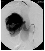
The initial therapy for treatment of arteriovenous malformations (AVMs) is usually angiographically guided embolization of the malformation. Numerous substances have been injected in an effort to ablate the arteriovenous malformation (AVM). Initial attempts at embolization were complicated by recurrence of the arteriovenous malformation (AVM). This was thought to be due to the type of material used for embolization. Materials that have been used for embolization include steel coils, autologous blood clots, gelatin sponges and foams, and synthetic polymers.
The most effective material for embolization appears to be absolute alcohol, which is relatively inexpensive. Injection through the catheter lumen is also easier than with many of the synthetic materials. Balloon catheters are used to occlude the feeding artery to prevent retrograde migration of the alcohol. The alcohol denatures the proteins within the wall of the arteriovenous malformation (AVM), thereby inducing thrombosis and occlusion of the malformations. Superselective embolization with coils and microspheres has also been described.
Repeat treatments may be needed to completely ablate the arteriovenous malformation (AVM). Alcohol or other material can be used for the subsequent treatments. Epinephrine injection before embolization may make the procedure more effective by inducing vasospasm, thereby concentrating the injected material within the arteriovenous malformation (AVM).
Surgical Therapy
The treatment most likely to cure an arteriovenous malformation (AVM) is total nephrectomy. Total nephrectomy is indicated for large cirsoid arteriovenous malformations (AVMs). In most cases, nephrectomy is reserved for patients in whom more conservative therapy has failed. If the fistula is due to malignancy, then radical nephrectomy is usually indicated.
The primary criticism of nephrectomy for renal arteriovenous malformations (AVMs) is that significant amounts of normal renal tissue are removed. Thus, reconstructive approaches have been advocated in selected circumstances. Partial nephrectomy has been accepted as a safe treatment for small, polar lesions. With increasing experience with partial nephrectomy for malignancy, partial nephrectomy will likely be attempted with greater confidence, even for large and centrally located arteriovenous malformations (AVMs). Additionally, to decrease the morbidity from the incisions needed for renal surgery, laparoscopic partial and total nephrectomy have been used with increasing frequency to treat selected renal arteriovenous malformations (AVMs).
In addition to partial nephrectomy, other techniques have been used to treat arteriovenous malformations (AVMs). Small malformations located in the peripheral aspect of the kidney may be treated by ligation of feeding vessels. The dissection of the feeding vessels may be technically difficult. Bench surgery with autotransplantation may facilitate the successful treatment of large and/or centrally located malformations. This degree of renal reconstruction is rarely necessary but may preserve enough functional renal tissue to avoid dialysis in select cases.
Despite being the most successful treatment for renal arteriovenous malformations (AVMs), surgical intervention is usually reserved for those cases refractory to embolization or those associated with malignancy.
Surgery India Renal Arteriovenous Malformation, Cost Renal Surgery, Renal Arteriovenous Malformation Treatment, Renal Arteriovenous Malformation, India Renal Arteriovenous Malformation Treatment Hospital, India Renal Arteriovenous Malformation Treatment, India Renal Av Malformation, Surgery India Tour, India Renal Arteriovenous Fistula, India Renal Tumor, India Renal Arteriovenous Malformation Surgery, India Arteriovenous Malformation, India Cost Renal Arteriovenous Malformation Treatment, India Renal, India Malformation, India Arteriovenous, India Low Cost Renal Arteriovenous Malformation Treatment Hospital